آزمیران
دستگاهای آزمایشگاهی
مواد شیمیایی
شیشه آلات
محیط کشتهای میکروبی
فیلتر یا کاغذ صافی
ملزومات
سکوبندی
مقالات و توضیحات
جستجوهای مرتبط
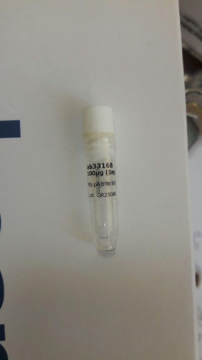
Anti-VE Cadherin antibody - Intercellular Junction Marker ab33168 100 µg
Anti-VE Cadherin antibody - Intercellular Junction Marker ab33168 100 µg
اعتبار قیمت 95.7.1
لطفا پس از پایان اعتبار قیمت با تلفن 88920648 تماس حاصل فرمائید
Overview
- Product nameAnti-VE Cadherin antibody - Intercellular Junction Marker
See all VE Cadherin primary antibodies - DescriptionRabbit polyclonal to VE Cadherin - Intercellular Junction Marker
- Tested applicationsICC/IF, WB, IHC-Fr, IP, In-Cell ELISA, IHC-P, Flow Cytmore details
- Species reactivityReacts with: Mouse, Chicken, Human
Predicted to work with: Cow, Pig
- Immunogen
Synthetic peptide conjugated to KLH derived from within residues 750 to the C-terminus of Human VE Cadherin.
(Peptide available as ab27462.)
- Positive control
- This antibody gave a positive signal in HUVEC (Human umbilical vein epithelial) Cell Lysate.
Properties
- FormLiquid
- Storage instructionsShipped at 4°C. Store at +4°C short term (1-2 weeks). Upon delivery aliquot. Store at -20°C or -80°C. Avoid freeze / thaw cycle.
- Storage bufferPreservative: 0.02% Sodium Azide
Constituents: 1% BSA, PBS, pH 7.4 -
Concentration 100 µg at 1 mg/ml
- PurityImmunogen affinity purified
- ClonalityPolyclonal
- IsotypeIgG
- Research areas
Applications
Our Abpromise guarantee covers the use of ab33168 in the following tested applications.
The application notes include recommended starting dilutions; optimal dilutions/concentrations should be determined by the end user.
| Application | Abreviews | Notes |
|---|---|---|
| ICC/IF | Use a concentration of 5 µg/ml. | |
| WB | Use a concentration of 1 µg/ml. Detects a band of approximately 115 kDa (predicted molecular weight: 87 kDa). | |
| IHC-Fr | Use at an assay dependent concentration. | |
| IP | Use at an assay dependent concentration. | |
| In-Cell ELISA | Use at an assay dependent concentration. PubMed: 22689949 | |
| IHC-P | Use at an assay dependent concentration. Perform heat mediated antigen retrieval with Tris/EDTA buffer pH 9.0 before commencing with IHC staining protocol. | |
| Flow Cyt | Use at an assay dependent concentration. ab171870-Rabbit polyclonal IgG, is suitable for use as an isotype control with this antibody. |
Target
- FunctionCadherins are calcium dependent cell adhesion proteins. They preferentially interact with themselves in a homophilic manner in connecting cells; cadherins may thus contribute to the sorting of heterogeneous cell types. This cadherin may play a important role in endothelial cell biology through control of the cohesion and organization of the intercellular junctions. It associates with alpha-catenin forming a link to the cytoskeleton.
- Tissue specificityEndothelial tissues and brain.
- Sequence similaritiesContains 5 cadherin domains.
- Post-translational
modificationsPhosphorylated on tyrosine residues by KDR/VEGFR-2. Dephosphorylated by PTPRB. - Cellular localizationCell junction. Cell membrane. Found at cell-cell boundaries and probably at cell-matrix boundaries.
- Information by UniProt
-
Database links
- Entrez Gene: 1003 Human
- Entrez Gene: 12562 Mouse
- Omim: 601120 Human
- SwissProt: P33151 Human
- SwissProt: P55284 Mouse
- Unigene: 76206 Human
- Unigene: 21767 Mouse
-
Alternative names
- 7B 4 antibody
- 7B4 antibody
- 7B4 antigen antibody
see all
Anti-VE Cadherin antibody - Intercellular Junction Marker images
-
 Immunocytochemistry/ Immunofluorescence - Anti-VE Cadherin antibody - Intercellular Junction Marker (ab33168)
ab33168 stained HUVEC cells. The cells were 100% methanol fixed for 5 minutes at -20°C and then incubated in 1%BSA / 10% normal goat serum / 0.3M glycine in 0.1% PBS-Tween for 1hour at room temperature to permeabilise the cells and block non-specific protein-protein interactions. The cells were then incubated with the antibody (ab33168 at 1µg/ml) overnight at +4°C. The secondary antibody (pseudo-colored green) was Goat Anti-Rabbit IgG H&L (Alexa Fluor® 488) preadsorbed (ab150081) used at a 1/1000 dilution for 1hour at room temperature. Alexa Fluor® 594 WGA was used to label plasma membranes (pseudo-colored red) at a 1/200 dilution for 1hour at room temperature. DAPI was used to stain the cell nuclei (pseudo-colored blue) at a concentration of 1.43µM for 1hour at room temperature.
-
 Immunocytochemistry/ Immunofluorescence - Anti-VE Cadherin antibody (ab33168)Stephen Yarwood, Inst Mol, Cell and Sys Bio, United KingdomICC/IF image of VE-Cadherin staining on HUVEC cells using ab33168. The cells were incubated with the primary antibody (ab33168) and the secondary was FITC conjugated anti-rabbit used at 1:400. The cells were incubated with only the secondary antibody as a negative control.
 -
 Immunocytochemistry/ Immunofluorescence - VE Cadherin antibody (ab33168)Ana Kasirer-Friede, Univ California-San Diego, Dept. Of Medicine,, United StatesICC/IF image of VE Cadherin stained HUVEC cells. The cells The cells were incubated with the antibody ab33168 at 1/150 (Green). The cells were also stained with Rhodamine phalloidin (Red).
 -
 Immunohistochemistry (Formalin/PFA-fixed paraffin-embedded sections) - VE Cadherin antibody (ab33168)This image is courtesy of an anonymous Abreviewab33168 (1/50) staining VE Cadherin in paraffin-embbeded mouse heart tissue sections. Tissue underwent fixation in formaldehyde, heat-mediated antigen retrieval in citrate buffer and blocking (5 minutes/peroxidase block and 10 minutes/protein block). For further experimental details please refer to abreview.
 -
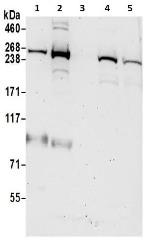
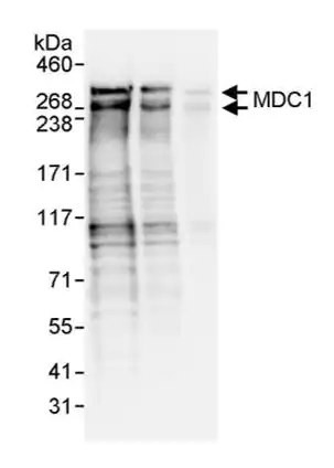
 Western blot - VE Cadherin antibody (ab33168)Anti-VE Cadherin antibody - Intercellular Junction Marker (ab33168) at 1 µg/ml + HUVEC Cell Lysate at 20 µg
Secondary
IRDye 680 Conjugated Goat Anti-Rabbit IgG (H+L) at 1/10000 dilution
Performed under reducing conditions.
Predicted band size : 87 kDa
Observed band size : 115 kDa (why is the actual band size different from the predicted?)
The band we observe at 115 kDa is believed to be the glycosylated form of the protein. -
 Flow Cytometry - VE Cadherin antibody (ab33168)This image is courtesy of an anonymous abreview.
ab33168 used in Flow cytometry.
Rabbit IgG isotype control (white)
Human REH B cells were fixed in paraformaldehyde and permeabilized using saponin. Primary antibody used undiluted (2µl in 100µl of cells in PBS) and incubated for 15 minutes at 4°C. The secondary antibody used was an undiluted, Alexa Fluor®488 conjugated goat anti-rabbit IgG. -
 Immunoprecipitation - Anti-VE Cadherin antibody - Intercellular Junction Marker (ab33168)This image is courtesy of an anonymous Abreview
ab33168 Immunoprecipitating VE Cadherin in human HUVEC whole cell lysate. 1000000 cells lysate was incubated with primary antibody (1/100 in 0.5% NP40, 150mM NaCl, 50mM Tris) and matrix (Dynabeads) for 2 hours at 4°C. For western blotting a HRP-conjugated mouse anti-VE Cadherin (1/3000) was used to confirm successful immunoprecipation.
References for Anti-VE Cadherin antibody - Intercellular Junction Marker (ab33168)
This product has been referenced in:
- Tsuneki M & Madri JA CD44 regulation of endothelial cell proliferation and apoptosis via modulation of CD31 and VE-cadherin expression. J Biol Chem 289:5357-70 (2014). Mouse . Read more (PubMed: 24425872) »
- Tata A et al. An image-based RNAi screen identifies SH3BP1 as a key effector of Semaphorin 3E-PlexinD1 signaling. J Cell Biol 205:573-590 (2014). Read more (PubMed: 24841563) »
فروشنده: آزمیران
گارانتی اصالت و سلامت کالا
پشتیبانی ۷ روز هفته ۲۴ ساعته
۲,۷۵۰,۰۰۰ تومان
محصولات مرتبط
:


![آنتی بادی ضد سیتوکراتین 7 [RCK105] - نشانگر اسکلت سلولی (ab9021) بسته بندی 100 میکروگرمی](/pics/38612_1669237731.jpg)
۶۹,۷۴۳,۰۴۱تومان
![آنتی بادی نوترکیب Anti-Ki67 [SP6] (ab16667) بسته بندی 100 میکرولیتری](/pics/38612_1670054494.jpg)
![آنتی بادی نوترکیب Anti-HIV1 p24 [P131] (ab32352) بسته بندی 100 میکرولیتر](/pics/38612_1669236804.jpg)
![آنتی بادی پلیمری ضد پلی (ADP-Ribose) [10H] (ab14459) بسته بندی 100 میکروگرم](/pics/38612_1669236588.jpg)
![آنتی بادی نوترکیب Anti-Rad51 [EPR4030(3)] (ab133534) بسته بندی 100 میکروگرم](/pics/38612_1669236410.jpg)
![آنتی بادی نوترکیب Anti-Nrf2 [EP1808Y] - درجه تراشه (ab62352) 100 میکرولیتر](/pics/38612_1669236150.jpg)
![آنتی بادی ضد CD130 (gp130) [B-S12] - بدون BSA و Azide100 میکروگرم (ab27359)](/pics/38612_1669235935.jpg)