آزمیران
دستگاهای آزمایشگاهی
مواد شیمیایی
شیشه آلات
محیط کشتهای میکروبی
فیلتر یا کاغذ صافی
ملزومات
سکوبندی
مقالات و توضیحات
جستجوهای مرتبط
![Anti-CREB (phospho S133) antibody [E113] ab32096 100 µl](/pic/p/200838/anti-creb-phospho-s133-antibody-e113-ab32096-100-%C2%B5l.jpeg)
Anti-CREB (phospho S133) antibody [E113] ab32096 100 µl
Anti-CREB (phospho S133) antibody [E113] ab32096 100 µl
اعتبار قیمت 95.7.1
لطفا پس از پایان اعتبار قیمت با تلفن 88920648 تماس حاصل فرمائید
Overview
- Product nameAnti-CREB (phospho S133) antibody [E113]
See all CREB primary antibodies - DescriptionRabbit monoclonal [E113] to CREB (phospho S133)
- SpecificityThis antibody is specific for CREB phosphorylated on Serine 133.
- Tested applicationsWB, IHC-P, IP, ICC/IFmore details
- Species reactivityReacts with: Mouse, Rat, Human
Predicted to work with: Chicken, Cow, Zebrafish
- Immunogen
Synthetic phoshopeptide corresponding to residues surrounding Serine 133 of human CREB.
- Positive control
- WB: A431 cell lysate. IF: A431 cells. IHC-P: Thyroid gland adenocarcinoma.
- General notes
Produced using Abcam’s RabMAb® technology. RabMAb® technology is covered by the following U.S. Patents, No. 5,675,063 and/or 7,429,487.
We are constantly working hard to ensure we provide our customers with best in class antibodies. As a result of this work we are pleased to now offer this antibody in purified format. We are in the process of updating our datasheets. The purified format is designated ‘PUR’ on our product labels. If you have any questions regarding this update, please contact our Scientific Support team.
Properties
- FormLiquid
- Storage instructionsShipped at 4°C. Store at +4°C short term (1-2 weeks). Upon delivery aliquot. Store at -20°C. Stable for 12 months at -20°C.
- Storage bufferpH: 7.20
Preservative: 0.01% Sodium azide
Constituents: 59% PBS, 40% Glycerol, 0.05% BSA -
Concentration Batch dependent within range: 100 µl at 0.695 - 0.696 mg/ml Batch dependent within range: 10 µl at 0.695 - 0.696 mg/ml
- PurityProtein A purified
- ClonalityMonoclonal
- Clone numberE113
- IsotypeIgG
- Research areas
Applications
Our Abpromise guarantee covers the use of ab32096 in the following tested applications.
The application notes include recommended starting dilutions; optimal dilutions/concentrations should be determined by the end user.
| Application | Abreviews | Notes |
|---|---|---|
| WB | 1/5000. Predicted molecular weight: 37 kDa.Can be blocked with CREB (phospho S133) peptide (ab182754). For unpurified use at 1/500. |
|
| IHC-P | 1/100. For unpurified use at 1/250. See protocols (link: http://www.abcam.com/protocols/ihc-antigen-retrieval-protocol).
|
|
| IP | 1/50. For unpurified use at 1/40. |
|
| ICC/IF | 1/100. For unpurified use at 1/250. |
Target
- FunctionThis protein binds the cAMP response element (CRE), a sequence present in many viral and cellular promoters. CREB stimulates transcription on binding to the CRE. Transcription activation is enhanced by the TORC coactivators which act independently of Ser-133 phosphorylation. Implicated in synchronization of circadian rhythmicity.
- Involvement in diseaseDefects in CREB1 may be a cause of angiomatoid fibrous histiocytoma (AFH) [MIM:612160]. A distinct variant of malignant fibrous histiocytoma that typically occurs in children and adolescents and is manifest by nodular subcutaneous growth. Characteristic microscopic features include lobulated sheets of histiocyte-like cells intimately associated with areas of hemorrhage and cystic pseudovascular spaces, as well as a striking cuffing of inflammatory cells, mimicking a lymph node metastasis. Note=A chromosomal aberration involving CREB1 is found in a patient with angiomatoid fibrous histiocytoma. Translocation t(2;22)(q33;q12) with CREB1 generates a EWSR1/CREB1 fusion gene that is most common genetic abnormality in this tumor type.
- Sequence similaritiesBelongs to the bZIP family.
Contains 1 bZIP domain.
Contains 1 KID (kinase-inducible) domain. - Post-translational
modificationsStimulated by phosphorylation. Phosphorylation of both Ser-133 and Ser-142 in the SCN regulates the activity of CREB and participates in circadian rhythm generation. Phosphorylation of Ser-133 allows CREBBP binding (By similarity). Phosphorylated upon DNA damage, probably by ATM or ATR.
Sumoylated by SUMO1. Sumoylation on Lys-304, but not on Lys-285, is required for nuclear localization of this protein. Sumoylation is enhanced under hypoxia, promoting nuclear localization and stabilization. - Cellular localizationNucleus.
- Information by UniProt
-
Database links
- Entrez Gene: 281713 Cow
- Entrez Gene: 1385 Human
- Entrez Gene: 12912 Mouse
- Entrez Gene: 81646 Rat
- Omim: 123810 Human
- SwissProt: P27925 Cow
- SwissProt: P16220 Human
- SwissProt: Q01147 Mouse
see all -
Alternative names
- Active transcription factor CREB antibody
- cAMP response element binding protein 1 antibody
- cAMP response element binding protein antibody
see all
Anti-CREB (phospho S133) antibody [E113] images
-
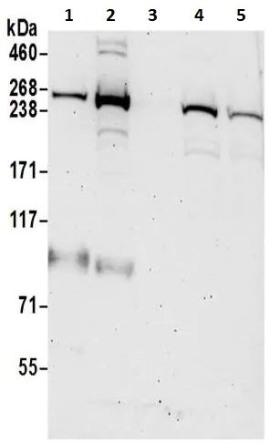
![Western blot - Anti-CREB (phospho S133) antibody [E113] (ab32096)](http://a.abcam.com/ps/products/32/ab32096/Images/ab32096-13-ab32096WB3.jpg) Western blot - Anti-CREB (phospho S133) antibody [E113] (ab32096)Lane 1 : Anti-CREB (phospho S133) antibody [E113] (ab32096) at 1/5000 dilution (purified)
Western blot - Anti-CREB (phospho S133) antibody [E113] (ab32096)Lane 1 : Anti-CREB (phospho S133) antibody [E113] (ab32096) at 1/5000 dilution (purified)
Lane 2 : purified at 1/5000 dilution
Lane 1 : Untreated C6 cell lysate
Lane 2 : C6 treated with Calyculin A cell lysate
Lysates/proteins at 10 µg per lane.
Secondary
Goat Anti-Rabbit IgG H&L (HRP) (ab97051) at 1/1000 dilution (Goat Anti-Rabbit IgG, (H+L), Peroxidase conjugated)
Predicted band size : 37 kDaBlocking buffer and concentration: 5% NFDM/TBST.
Diluting buffer and concentration: 5% NFDM /TBST. -
![Western blot - Anti-CREB (phospho S133) antibody [E113] (ab32096)](http://a.abcam.com/ps/products/32/ab32096/Images/ab32096-12-ab32096WB2.jpg) Western blot - Anti-CREB (phospho S133) antibody [E113] (ab32096)All lanes : Anti-CREB (phospho S133) antibody [E113] (ab32096) at 1/5000 dilution (purified)
Western blot - Anti-CREB (phospho S133) antibody [E113] (ab32096)All lanes : Anti-CREB (phospho S133) antibody [E113] (ab32096) at 1/5000 dilution (purified)
Lane 1 : Untreated NIH/3T3 cell lysate
Lane 2 : NIH/3T3 treated with Calyculin A
Lysates/proteins at 10 µg per lane.
Secondary
Goat Anti-Rabbit IgG H&L (HRP) (ab97051) at 1/1000 dilution (Goat Anti-Rabbit IgG, (H+L), Peroxidase conjugated)
Predicted band size : 37 kDaBlocking buffer and concentration: 5% NFDM/TBST.
Diluting buffer and concentration: 5% NFDM /TBST. -
![Western blot - Anti-CREB (phospho S133) antibody [E113] (ab32096)](http://a.abcam.com/ps/products/32/ab32096/Images/ab32096-11-ab32096WB1.jpg) Western blot - Anti-CREB (phospho S133) antibody [E113] (ab32096)All lanes : Anti-CREB (phospho S133) antibody [E113] (ab32096) at 1/5000 dilution (purified)
Western blot - Anti-CREB (phospho S133) antibody [E113] (ab32096)All lanes : Anti-CREB (phospho S133) antibody [E113] (ab32096) at 1/5000 dilution (purified)
Lane 1 : Untreated HeLa cell lysate
Lane 2 : HeLa treated with Calyculin A cell lysate
Lysates/proteins at 10 µg per lane.
Secondary
Goat Anti-Rabbit IgG H&L (HRP) (ab97051) at 1/1000 dilution (Goat Anti-Rabbit IgG, (H+L), Peroxidase conjugated)
Predicted band size : 37 kDaBlocking buffer and concentration: 5% NFDM/TBST.
Diluting buffer and concentration: 5% NFDM /TBST. -
![Western blot - Anti-CREB (phospho S133) antibody [E113] (ab32096)](http://a.abcam.com/ps/datasheet/images/32/ab32096/CREB-Primary-antibodies-ab32096-4.jpg) Western blot - Anti-CREB (phospho S133) antibody [E113] (ab32096)
Western blot - Anti-CREB (phospho S133) antibody [E113] (ab32096)
developed using the ECL technique
Performed under reducing conditions.
Predicted band size : 37 kDaSK-N-SH cells were incubated at 37°C for 30 minutes with vehicle control (0 µM) and different concentrations of (R,S)-CHPG (ab120039). Increased expression of CREB (phospho S133) in SK-N-SH cells correlates with an increase in (R,S)-CHPG concentration, as described in literature.
Whole cell lysates were prepared with RIPA buffer (containing protease inhibitors and sodium orthovanadate), 20µg of each were loaded on the gel and the WB was run under reducing conditions. After transfer the membrane was blocked for an hour using 3% milk before being incubated with unpurified ab32096 at 1/500 dilution and ab32515 at 1 µg/ml overnight at 4°C. Antibody binding was detected using an anti-rabbit antibody conjugated to HRP (ab97051 ) at 1/10000 dilution and visualised using ECL development solution.
-
![Western blot - Anti-CREB (phospho S133) antibody [E113] (ab32096)](http://a.abcam.com/ps/datasheet/images/32/ab32096/CREB-Primary-antibodies-ab32096-5.jpg) Western blot - Anti-CREB (phospho S133) antibody [E113] (ab32096)
Western blot - Anti-CREB (phospho S133) antibody [E113] (ab32096)
Predicted band size : 37 kDaSK-N-SH cells were incubated at 37°C for 30 minutes with vehicle control (0 µM) and different concentrations of (S)-3,5-DHPG (ab120007). Increased expression of CREB (phospho S133) in SK-N-SH cells correlates with an increase in (S)-3,5-DHPGconcentration, as described in literature.
Whole cell lysates were prepared with RIPA buffer (containing protease inhibitors and sodium orthovanadate), 20µg of each were loaded on the gel and the WB was run under reducing conditions. After transfer the membrane was blocked for an hour using 3% milk before being incubated with unpurified ab32096 at 1/500 dilution and ab32515 at 1 µg/ml overnight at 4°C. Antibody binding was detected using an anti-rabbit antibody conjugated to HRP (ab97051) at 1/10000 dilution and visualised using ECL development solution.
-
![Immunoprecipitation - Anti-CREB (phospho S133) antibody [E113] (ab32096)](http://a.abcam.com/ps/products/32/ab32096/Images/ab32096-10-ab32096IP1.jpg) Immunoprecipitation - Anti-CREB (phospho S133) antibody [E113] (ab32096)
Immunoprecipitation - Anti-CREB (phospho S133) antibody [E113] (ab32096)ab32096 (purified) at 1/50 immunoprecipitating CREB (phospho S133) in HeLa whole cell lysate. 10 ug of cell lysate was present in the input. For western blotting, a HRP-conjugated Veriblot for IP secondary antibody (ab131366) (1/1,500) was used as the secondary antibody. A rabbit monoclonal IgG (ab172730) was used intead of ab128913 as a negative control (Lane 3).
Blocking buffer and concentration: 5% NFDM/TBST.
Diluting buffer and concentration: 5% NFDM /TBST.
-
![Immunohistochemistry (Formalin/PFA-fixed paraffin-embedded sections) - Anti-CREB (phospho S133) antibody [E113] (ab32096)](http://a.abcam.com/ps/products/32/ab32096/Images/ab32096-8-ab32096IHC1.jpg) Immunohistochemistry (Formalin/PFA-fixed paraffin-embedded sections) - Anti-CREB (phospho S133) antibody [E113] (ab32096)Immunohistochemistry (Formalin/PFA-fixed paraffin-embedded sections) analysis of human thyroid carcinoma tissue labelling CREB with purified ab32096 at 1/100. Heat mediated antigen retrieval was performed using Tris/EDTA buffer pH 9. ab97051, a goat anti-rabbit IgG H&L (HRP) was used as the secondary antibody (1/500). Negative control using PBS instead of primary antibody. Counterstained with hematoxylin.
Immunohistochemistry (Formalin/PFA-fixed paraffin-embedded sections) - Anti-CREB (phospho S133) antibody [E113] (ab32096)Immunohistochemistry (Formalin/PFA-fixed paraffin-embedded sections) analysis of human thyroid carcinoma tissue labelling CREB with purified ab32096 at 1/100. Heat mediated antigen retrieval was performed using Tris/EDTA buffer pH 9. ab97051, a goat anti-rabbit IgG H&L (HRP) was used as the secondary antibody (1/500). Negative control using PBS instead of primary antibody. Counterstained with hematoxylin. -
![Immunohistochemistry (Formalin/PFA-fixed paraffin-embedded sections) - Anti-CREB (phospho S133) antibody [E113] (ab32096)](http://a.abcam.com/ps/datasheet/Images/32/ab32096/ab32089_3.jpg) Immunohistochemistry (Formalin/PFA-fixed paraffin-embedded sections) - Anti-CREB (phospho S133) antibody [E113] (ab32096)
Immunohistochemistry (Formalin/PFA-fixed paraffin-embedded sections) - Anti-CREB (phospho S133) antibody [E113] (ab32096)Immunohistochemistry (Formalin/PFA-fixed paraffin-embedded sections) analysis of human thyroid gland adenocarcinoma tissue labelling CREB with unpurified ab32096 at 1/250 dilution.
Panel A: Cells are untreated.
Panel B: Cells are treated with Phosphatase. -
![Immunohistochemistry (Formalin/PFA-fixed paraffin-embedded sections) - Anti-CREB (phospho S133) antibody [E113] (ab32096)](http://a.abcam.com/ps/datasheet/images/32/ab32096/CREB-Primary-antibodies-ab32096-3.jpg) Immunohistochemistry (Formalin/PFA-fixed paraffin-embedded sections) - Anti-CREB (phospho S133) antibody [E113] (ab32096)This image is courtesy of an Abreivew submitted by Akiko Shingo.
Immunohistochemistry (Formalin/PFA-fixed paraffin-embedded sections) - Anti-CREB (phospho S133) antibody [E113] (ab32096)This image is courtesy of an Abreivew submitted by Akiko Shingo.Immunohistochemistry (Formalin/PFA-fixed paraffin-embedded sections) analysis of rat hippocampus tissue labelling CREB with unpurified ab32096 at 1/100 dilution. Sections were subjected to antigen retrieval by autoclave prior to blocking with 8% milk for 30 minutes at 37°C. The primary antibody was diluted 1/100 with DAKO antibody diluent and incubated with the sample for 18 hours at 4°C. An LSAB-labeled Streptavidin-Biotin conjugated Goat polyclonal antibody was used undiluted as the secondary antibody.
-
![Immunocytochemistry/ Immunofluorescence - Anti-CREB (phospho S133) antibody [E113] (ab32096)](http://a.abcam.com/ps/products/32/ab32096/Images/ab32096-9-ab32096ICCIF1.jpg) Immunocytochemistry/ Immunofluorescence - Anti-CREB (phospho S133) antibody [E113] (ab32096)
Immunocytochemistry/ Immunofluorescence - Anti-CREB (phospho S133) antibody [E113] (ab32096)Immunocytochemistry/Immunofluorescence analysis of A431(human epidermoid carcinoma) cells +/- AP 37℃ 1h labelling CREB with purified ab32096 at 1/100. Cells were fixed with 4% paraformaldehyde and permeabilized with 0.1% Triton X-100. ab150077, an Alexa Fluor® 488-conjugated goat anti-rabbit IgG (1/500) was used as the secondary antibody. DAPI (blue) was used as the nuclear counterstain. ab7291, a mouse anti-tubulin (1/1000) and ab150120, an Alexa Fluor® 594-conjugated goat anti-mouse IgG (1/1000) were also used.
Control 1: primary antibody (1/100) and secondary antibody, ab150120, an Alexa Fluor® 594-conjugated goat anti-mouse IgG (1/500).
Control 2: ab7291 (1/1000) and secondary antibody, ab150077, an Alexa Fluor® 488-conjugated goat anti-rabbit IgG (1/500).
-
![Immunocytochemistry/ Immunofluorescence - Anti-CREB (phospho S133) antibody [E113] (ab32096)](http://a.abcam.com/ps/datasheet/Images/32/ab32096/ab32089_2.jpg) Immunocytochemistry/ Immunofluorescence - Anti-CREB (phospho S133) antibody [E113] (ab32096)
Immunocytochemistry/ Immunofluorescence - Anti-CREB (phospho S133) antibody [E113] (ab32096)Immunocytochemistry/Immunofluorescence analysis of A431 cells labelling CREB with unpurified ab32096 at 1/250.
Panel A: Cells are untreated.
Panel B: Cells are treated with Phosphatase.
References for Anti-CREB (phospho S133) antibody [E113] (ab32096)
This product has been referenced in:
- Liu F et al. The oncoprotein HBXIP enhances angiogenesis and growth of breast cancer through modulating FGF8 and VEGF. Carcinogenesis 35:1144-53 (2014). Read more (PubMed: 24464787) »
- Liu L et al. Genetic deletion of calcium/calmodulin-dependent protein kinase kinase ß (CaMKK ß) or CaMK IV exacerbates stroke outcomes in ovariectomized (OVXed) female mice. BMC Neurosci 15:118 (2014). WB ; Mouse . Read more (PubMed: 25331941) »
فروشنده: آزمیران
گارانتی اصالت و سلامت کالا
پشتیبانی ۷ روز هفته ۲۴ ساعته
۲,۷۵۰,۰۰۰ تومان
محصولات مرتبط
:


![آنتی بادی ضد سیتوکراتین 7 [RCK105] - نشانگر اسکلت سلولی (ab9021) بسته بندی 100 میکروگرمی](/pics/38612_1669237731.jpg)
۶۹,۷۴۳,۰۴۱تومان
![آنتی بادی نوترکیب Anti-Ki67 [SP6] (ab16667) بسته بندی 100 میکرولیتری](/pics/38612_1670054494.jpg)
![آنتی بادی نوترکیب Anti-HIV1 p24 [P131] (ab32352) بسته بندی 100 میکرولیتر](/pics/38612_1669236804.jpg)
![آنتی بادی پلیمری ضد پلی (ADP-Ribose) [10H] (ab14459) بسته بندی 100 میکروگرم](/pics/38612_1669236588.jpg)
![آنتی بادی نوترکیب Anti-Rad51 [EPR4030(3)] (ab133534) بسته بندی 100 میکروگرم](/pics/38612_1669236410.jpg)
![آنتی بادی نوترکیب Anti-Nrf2 [EP1808Y] - درجه تراشه (ab62352) 100 میکرولیتر](/pics/38612_1669236150.jpg)
![آنتی بادی ضد CD130 (gp130) [B-S12] - بدون BSA و Azide100 میکروگرم (ab27359)](/pics/38612_1669235935.jpg)