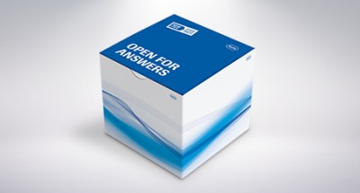
The High Pure PCR Template Preparation Kit purifies nucleic acids from a
wide variety of sample materials, including whole blood, cultured
cells, and tissue samples. Bacteria and yeast require a specific
pre-lysis treatment with lysozyme or lyticase.
- Minimize DNA loss using a kit that removes contaminants without precipitation or other handling steps that degrade DNA.
- Recover high molecular weight DNA (30 to 50 kb) that is suitable for long-template PCR.
- Improve reliability and reproducibility in downstream applications (real-time, quantitative PCR).
- Save time and maximize flexibility by preparing multiple PCR templates simultaneously.
- Eliminate the use of hazardous organic compounds such as cesium chloride, phenol, chloroform, and ethidium bromide.
The High Pure PCR Template Preparation Kit purifies nucleic acids from a
wide variety of sample materials including whole blood, cultured cells,
and tissue. The isolated nucleic acids can be used for:
- Long-template PCR (see Figure 1)
- Real-time, quantitative PCR
- SNP detection
- Southern blotting (see Figure 2)
- Cloning
> PCR amplification of long templates from nucleic acid samples
> Southern blot analysis of nucleic acid samples
> PCR amplification of nucleic acids prepared from paraffin-embedded tissue
> Analysis of PCR products specific for S. aureus (left) and various lactobacilli
Figure 1: PCR amplification of long templates from nucleic acid samples obtained with the High Pure PCR Template Preparation Kit.
Nucleic acids were prepared from blood or cultured human K562 cells. 250 ng of each preparation was amplified using a tissue-plasminogen-activator (tPA)-specific primer and either the Expand Long Template PCR System or the Expand 20 kbPLUS PCR System.

Lane 2 : 6.3 kb; obtained from blood and amplified using Expand Long Template PCR System buffer 1.
Lane 3 : 15 kb; obtained from blood and amplified using Expand Long Template PCR System buffer 3.
Lane 4 : 23 kb; obtained from blood and amplified using Expand Long Template PCR System buffer 3.
Lane 5 : 28 kb; obtained from K562 cells and amplified using Expand 20 kbPLUS PCR System.
Result: All High Pure preparations contained genomic DNA that yielded a distinct band of the expected size.
Figure 2: Southern blot analysis of nucleic acid samples obtained with the High Pure PCR Template Preparation Kit.
Nucleic acids were prepared from cultured mammalian cells (106 K562 cells) and from calf thymus tissue (25 mg). Aliquots of the preparations were digested with Eco RI, purified (see below), separated electrophoretically, and transferred to a membrane by Southern blotting. The blot was hybridized to a DIG-labeled beta-actin antisense RNA probe according to a standard procedure. The hybridized bands (i.e., beta-actin sequences) were detected with an alkaline phosphatase-labeled anti-DIG antibody and visualized via chemiluminescence with CSPD substrate. The blot was exposed to X-ray film for 1 hour.

Lanes 1, 9: DNA Molecular Weight Marker III.
Lanes 2, 3: 1 µg and 3 µg samples of K562 genomic DNA, purified with the High Pure PCR Product Purification Kit after Eco RI digestion.
Lanes 4-6: 0.3 µg, 1 µg, and 3 µg samples of K562 genomic DNA, purified by conventional phenol/chloroform extraction and ethanol precipitation after Eco RI digestion.
Lanes 7, 8: 0.4 µg and 4 µg samples of calf thymus genomic DNA, purified by conventional phenol/chloroform extraction and ethanol precipitation after Eco RI digestion
Result: All genomic DNA samples isolated using the High Pure PCR Template Preparation Kit were readily and completely cut by Eco RI, were suitable for Southern blot analysis, and contained the expected small and large fragments of the actin gene.
Figure 3: PCR amplification of nucleic acids prepared from paraffin-embedded tissue with the High Pure PCR Template Preparation Kit.
Nucleic acids were purified from formalin-fixed, paraffin-embedded samples of human prostate tissue. An aliquot (10 µL) of each nucleic acid product was used as template for the Expand High Fidelity PCR System (100 µL reaction mixture). The primers were derived from exon 7 of the androgen receptor gene. Aliquots (20 µL) of each amplicon were separated electrophoretically and the gel was stained with ethidium bromide.

Lane 1: DNA Molecular Weight Marker VIII.
Lane 2: Androgen receptor amplicon from a full tissue section.
Lane 3: Androgen receptor amplicon from half of a full tissue section.
Result: The 263 bp androgen receptor amplicon was clearly visible in both samples.
Figure 4: Analysis of PCR products specific for S. aureus (left) and various lactobacilli (right).
PCR templates were prepared from various samples (prepared as described below) with the High Pure PCR Template Preparation Kit, and analyzed by agarose gel electrophoresis.

Left panel
Lanes 1,12: DNA Molecular Weight Marker, 1 kb ladder.
Lane 2: Negative control (no DNA).
Lanes 3-5: S. aureus; lysis without lysostaphin; incubation periods of 15, 30, and 60 minutes.
Lanes 6-8: S. aureus; lysis with 10 µ/mL lysostaphin; incubation periods of 15, 30, and 60 minutes.
Lanes 9-11: S. aureus; lysis with 25 µ/mL lysostaphin; incubation periods of 15, 30, and 60 minutes.
Right panel
Lanes 13,18: DNA Molecular Weight Marker, phage lambda DNA, Hind III digest.
Lane 14: L. casei
Lane 15: L. curvatus
Lane 16: L. sakei
Lane 17: Negative control (no DNA).
Result [expressed as DNA concentration (µg/ml) in eluates]:
Incubation Period
15 min 30 min 60 min
L. casei
Without Mu 15.2 (40.0 %) 20.2 (53.2 %) 21.8 (57.4 %)
0.25 U/µL Mu 15.3 (40.3 %) 24.6 (64.7 %) 24.0 (63.2 %)
0.5 U/µL Mu 16.4 (43.2 %) 38.0 (100 %) 30.4 (80.0 %)
L. curvatus
Without Mu 9.2 (10.6 %) 14.7 (17.0 %) 26.1 (30.2 %)
0.25 U/µL Mu 25.9 (29.9 %) 61.3 (70.9 %) 70.7 (81.7 %)
0.5 U/µL Mu 46.0 (53.2 %) 86.5 (100 %) 131.1 (151.6 %)
L. sakei
Without Mu 13.5 (11.4 %) 16.6 (14.0 %) 52.9 (44.7 %)
0.25 U/µL Mu 35.0 (29.6 %) 71.8 (60.6 %) 168.2 (142.1 %)
0.5 U/µL Mu 79.3 (67.0 %) 118.4 (100 %) 196.1 (165.6 %)
S. aureus
Without Ly 10.3 (10.8 %) 30.5 (31.9 %) 64.2 (67.2 %)
10 µg/mL Ly 16.5 (17.3 %) 95.0 (99.5 %) 105.2 (110.2 %)
25 µg/mL Ly 16.2 (17.0 %) 95.5 (100 %) 106.7 (111.7 %)
Note: The lysis buffer contained 10 mg/mL lysozyme for lactobacilli and 1 mg/mL lysozyme for staphylococci together with the amounts of mutanolysin (MU) or lysostaphin (Ly) shown in the first column. The percentages refer to the DNA yield obtained with the highest mutanolysin or lysostaphin concentration and a 30 minute incubation period.
Nucleic acids bind to the surface of the glass fiber fleece in the
presence of a chaotropic salt (guanidine HCl). This allows the High
Pure filter tube to specifically immobilize nucleic acids (both DNA and
RNA) while they are freed of contaminants.
Note: A special Inhibitor Removal Buffer is
included which allows the use of heparinized sample material (100 U/mL
of heparin). This buffer increases the sensitivity and reproducibility
of assays performed with the isolated nucleic acid, even when the sample
contains heparin.
Capacity: The High Pure Spin Filter Tubes hold up to 700 μL sample volume.
Typical DNA Recovery
|
Starting Material and Quantity |
Yield/Recovery |
Time Required |
Number of Reactions |
| Mammalian whole blood, 200 – 300 µl | 3 – 9 µg | 20 minutes | 100/200 µL |
| Cultured mammalian cells, 104 – 108 | 15 – 20 µg | 20 minutes | |
| Mammalian solid tissue, 25 – 50 mg | 5 – 20 µg | 80 minutes, including lysis | |
| Mouse tail, 0.2 – 0.5 cm (25 – 50 mg) | 5 – 10 µg | 200 minutes, including lysis | |
| Yeast, 108 cells | 10 – 13 µg | 50 minutes, including lyticase digest | |
| Bacteria (gram positive or gram negative), 109 cells | 1 – 3 µg | 35 minutes, including lysozyme digest | |
| Dried blood spots | Product detectable by PCR | 80 minutes, including lysis | |
| buffy coat, 200 μl | 20 µg |
Principle
Blood cells or tissue are lysed by a short incubation with a special
Lysis Buffer and Proteinase K in the presence of a chaotropic salt such
as guanidine-HCl, which immediately inactivates all nucleases. Cellular
nucleic acids bind selectively to special glass fibers pre-packed in the
High Pure Purification Filter Tube. Bound nucleic acid is purified in a
series of rapid wash-and-spin steps to remove contaminating cellular
components. Finally, low salt elution releases the nucleic acid from the
glass fiber. This simple method eliminates the need for organic solvent
extractions and DNA precipitation, allowing for rapid purification of
many samples simultaneously.

Quality
DNA is isolated from 25 mg of calf thymus, 1 x 10⁶ K562 cells, and 200 µl of EDTA whole blood. Yield is measured via OD for tissue and cell samples. The quality of the nucleic acid is controlled in an Expand Long Range PCR with a 9.3 kb amplification product for DNA derived from cells. PCR on a LightCycler® Instrument is performed on human blood research samples using kits for Factor V Leiden and CycA.
Alternate DE MSDS - Material Safety Data Sheet
Alternate DE Instructions for Use - Version 22
Select the Version Number printed on the outer label of the product box.
To select documents, click on the download button shown beside.
Rapid and Reliable Methylation Detection in Archival Tissue Samples using High-Resolution Melting Analysis
اعتبار قیمت 97.1.1
